Volume EM

Selected as a method of the year by Nature’s Technologies [1] - the VolumeEM method aims to deliver electron microscopy resolution in the volumetric landscape, elucidating the three-dimensional organization of material and biological specimens at the nanoscale level. VolumeEM unit of the TUM Electron Microscopy Facility contributes to strengthening of TUM as a prime site for advanced electron microscopy methods, helping researchers from the fields of life sciences, microelectronics, and material science to answer their scientific questions.
The core instrument of the unit is a state-of-the-art dual-beam Crossbeam 550 electron microscope (Carl Zeiss, Germany), equipped with a windowless Ultim Extreme EDX detector (Oxford Instruments, United Kingdom) and a set of auxiliaries, including retractable low-voltage detector, gas injection systems, micromanipulator.
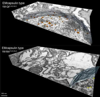
A unique area of TUM’s VolumeEM unit competence - is the application of genetically-encodable electron microscopy reporters for life science research under the supervision of Prof. Dr. Gil Westmeyer. Combined with the capacity to execute the full VolumeEM workflow in-house - this distinctive area of expertise aims to cover the BioEM community’s need for convenient and reliable EM reporters.
Next electron microscopy techniques are available in the VolumeEM unit of the TUM Electron Microscopy Facility:
High-resolution 2D scanning electron microscopy;
Energy Dispersive X-ray spectroscopy;
Array tomography for Volume EM;
FIB-SEM for high-resolution Volume EM;
FIB-SEM for S/TEM lamella preparation.
HIGGINBO, AVID. "SEVEN TECHNOLOGIES TO WATCH IN 2023." Nature 613 (2023): 26.