MTC1.2 MicroCT & X-ray phase contrasting imaging
Key instruments:

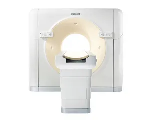
Clinical X-ray computed tomography scanner. Available equipment: X-ray tube (W-rotation anodes on a rotating gantry, max 120 kVp), motorized patient bed, image reconstruction software by Philips, powerful workstation for data processing
Typical applications: 3D visualization, tumor detection, scanning time of 2-10 s and an achievable resolution of 100 micrometers
| Principal investigator | Prof. Dr. Julia Herzen |
| PI's field of research | diagnostic X-ray imaging, X-ray instrumentation |
| Contact person | Kirsten Taphorn |
| Tel. | +49 89 289 12562 |
| kirsten.taphorn@tum.de | |
| Availability | cooperation |
| Lead time | ca. 1 week |
Instrument: X-ray Phasecontrast-Biopsyscanner

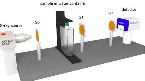
X-ray imaging instrument for biological samples. Available equipment: X-ray tube (Mo-rotating anode, max. 50 kVp), motorized sample rotation stage, set of optical transmission gratings for phase-contrast imaging, various detectors (flat panels, direct converting and energy resolving), image processing and reconstruction software, powerful workstation for data processing
Typical applications: 3D soft-tissue visualization, tumor detection, virtual histology; achievable spatial resolution: 40 micrometers
| Principal investigator | Prof. Dr. Julia Herzen |
| PI's field of research | X-ray phase contrast imaging, 3D virtual histology |
| Contact person | Kirsten Taphorn |
| Tel. | +49 89 289 12562 |
| kirsten.taphorn@tum.de | |
| Availability | cooperation |
| Lead time | ca. 1-2 weeks |
Instrument: GE Phoenix VtomeX

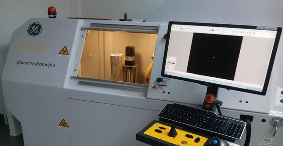
Medium-resolution tomographic X-ray imaging instrument for non-destructive testing and material science samples with a spatial resolution of < 2 micron, sample sizes < 30 cm and a maximum X-ray energy of 200 kVp; additional image reconstruction, image post-processing and 3D image visualisation software is available
Typical applications: non-destructive testing and material science samples
| Principal investigator | Prof. Dr. Franz Pfeiffer |
| PI's field of research | X-ray microscopy, Biomedical X-ray imaging, Artificial Intelligence & Biomedical imaging |
| Contact person | Dr. Klaus Achterhold |
| Tel. | +49 89 289 12559 |
| klaus.achterhold@tum.de | |
| Availability | cooperation |
| Lead time | ca. 1-2 weeks |
Instrument: Zeiss Xradia 520 Versa

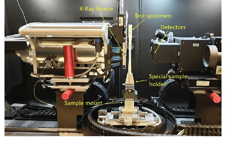
High-resolution tomographic X-ray imaging instrument for life science samples, biomaterials and biopsies with a spatial resolution of < 0.5 micron, sample sizes < 5 cm and a maximum X-ray energy of 180 kVp; additional image reconstruction, image post-processing and 3D image visualisation software is available
Typical applications: life science samples, biomaterials, biopsies
| Principal investigator | Prof. Dr. Franz Pfeiffer |
| PI's field of research | X-ray microscopy, Biomedical X-ray imaging, Artificial Intelligence & Biomedical imaging |
| Contact person | Dr. Klaus Achterhold |
| Tel. | +49 89 289 12559 |
| klaus.achterhold@tum.de | |
| Availability | cooperation |
| Lead time | ca. 1-2 weeks |
Instrument: FTIR with Focal Plane Array Detector
High-end mid-IR FTIR spectrometer (resolution 0.2 cm-1) with sample holders to work in transmission and ATR configuration, rapid scan option, polarization modulation and 2D FT-IR mapping capabilities
Typical applications: identification of both organic and inorganic compounds, detection of adsorbates and intermediates at electrochemical interfaces.
| Principal investigator | Prof. Dr. Katharina Krischer |
| PI's field of research | Self-Organization at Interfaces, Artificial Photosynthesis, Electrocatalysis |
| Contact person | Simon Stork |
| Tel. | +49 89 289 13877 |
| simon.stork@tum.de | |
| Availability | cooperation & pay-by-use |
| Lead time | depending on application |